
How the Human Eye Works
Human Eye: Anatomy, Structure & Its Functions
The human eye has been called the most complex organ in our body. It is amazing that so small can have so many parts. Eye is a natural optical instrument. It is present in the form of eyeball in the sockets of our skull. Its diameter is approximately 2.5 cm.
The human eye is an organ that reacts to light and allows vision. Rod and cone cells in the retina allow conscious light perception and vision including color differentiation and the perception of depth. The human eye can differentiate between about 10 million colors and is possibly capable of detecting a single photon. The eye is part of the sensory nervous system.
Similar to the eyes of other mammals, the human eye’s non-image-forming photosensitive ganglion cells in the retina receive light signals which affect adjustment of the size of the pupil, regulation and suppression of the hormone melatonin and entrainment of the body clock.
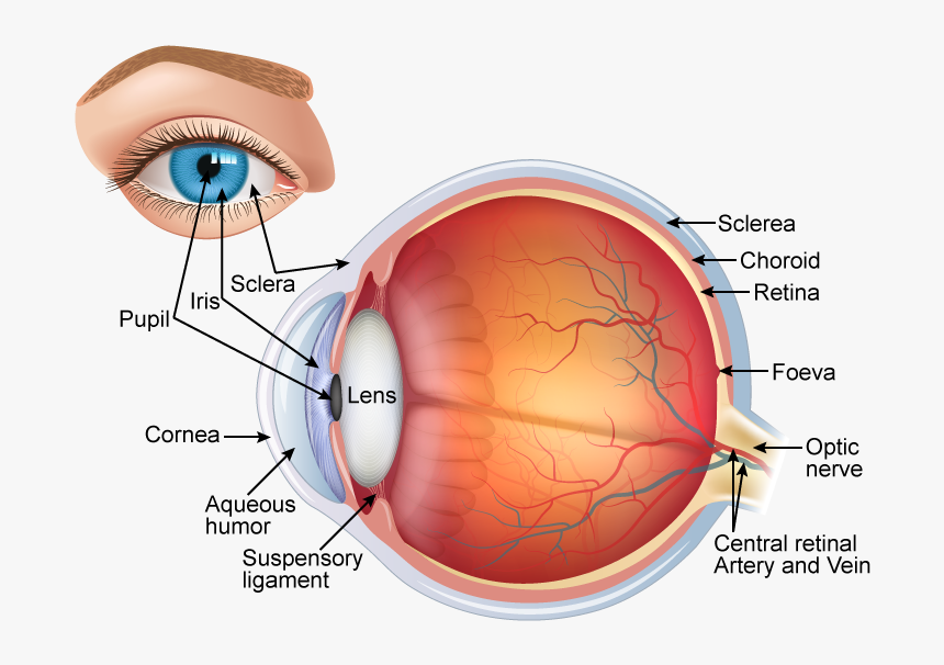
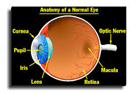
Anatomical structure of Eye
Eye ball consists of three layers
- Outer fibrous layer: Sclera, Cornea and Conjunctiva
- Middle vascular layer: Ciliary body, Choroid and Iris
- Inner layer: Retina
I. Outer fibrous layer:
It consists of following parts.
1. Sclera:
- It is the outermost, white, tough layer. It covers the whole part of the eyeball. It protects the eyeball. The front portion of the sclera is known as cornea.
- It covers 5/6 parts of eye ball.
- It maintains the shape of eye and provide attachment to the extrinsic muscle of eye
2. Cornea:
- It is a thin transparent front part of sclera.
- It forms a slight bulge at the front and covers an anterior 1/6 part of sclera.
- Cornea is avascular and absorbs oxygen from air.
- It refracts light to focus on retina.
- Cornea is covered by a thin, transparent membrane known as conjunctiva. Conjunctiva helps the eye to be moist and prevent the dryness.
3. Conjunctiva:
- It is a thin transparent layer that covers the cornea.
- It is formed of single layer of stratified squamous epithelium
- It protects the cornea.
II. Middle vascular layer:
It consists of following parts:
1. Choroid:
- It is thick vascular and pigmented layer situated below sclera.
- The pigmented cells absorbs light and prevent it from being reflected.
- The function of choroid is to provide nutrition and to prevent reflection of light.
2. Ciliary body:
- These are attached to choroid and present at the junction of sclera and cornea.
- It consists of two sets of ciliary muscle and suspensory ligament.
- Ciliary body is attached to lens and holds it in position
- Its function is to change the shape of lens by contraction or relaxation of muscle
3. Iris:
- It is muscular, pigmented and opaque diaphragm which hangs in the eye ball in front of lens.
- It has small circular opening called pupil.
- It has two types of muscles; circular and radial muscle. The movement of these muscles control the size of pupil.
- Pigment in iris gives color to eye.
- Iris control the amount of light entering into eye by controlling the size of pupil.
III. Inner layer:
It consists of photoreceptor cells and photo sensitive elements.
1. Retina:
- Retina is innermost layer.
- Neuroretina contains highly specialized photoreceptor nerve cells; rods and cones
- Each eye ball has 125 millions of rod cells and 7 millions of cone cells.
- Small depression in retinal wall is called Fovea centralis which contains only cone cells.
- Fovea centralis is highly sensitive to light and forms magnified image and give sharp and acute vision.
- The optic nerve enter retina at a point called blind spot. It does not contains any rods or cone cells. It is least sensitive to light and forms no image when light falls on blind spot
Rod cell:
- rods are sensors for perception of black to white shades
- Night vision is almost rod vision.
- It function in dim light
- Contains a photosensitive pigment rhodopsin formed from vitamin A.
Cone cell:
- Cones are sensors for perception of colors.
- It functions in bright light and differentiate colors.
- Contains a photosensitive pigment iodopsin.
Eye lens and chambers
1. Eye Lens:
- It is a large, flexible, transparent biconvex and fibrous crystalline body situated behind iris.
- Lens is enclosed in a transparent elastic capsule.
- Ciliary muscles control the thickness of lens and its power of accommodation.
- It forms the image of the object on retina.
- Lens separates the eye ball into two chamber
i. Aqueous chamber
ii. Vitreous chamber
Aqueous chamber:
- It is a smaller fluid filled chamber between cornea and lens.
- It is filled with aqueous humour containing aminoacids, glucose, ascorbic acid, hyaluronic acid and respiratory gases.
- The aqueous humour nourishes the lens and cornea and refracts light rays to focus on retina.
Vitreous chamber:
- It is a larger fluid filled chamber between lens and retina.
- It is filled with gelatinous vitreous humour containing salts and muco proteins
- It supports retina and refracts light to focus on retina.
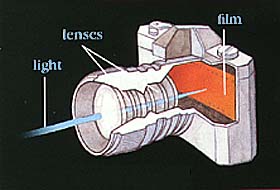
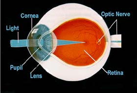
When light enter into the eye it passes through pupil. Iris controls the amount of the light. The cilliary muscles help the lens to focus the object on the retina. When light strikes on either on the rods or the cones of the retina, it is converted into an electric signal. The photoreceptors in the retina convert the light into the electrical signals. These signals carried to the brain by optic nerves and nerve fibers. The brain then translates the electrical signal into images we see.
Each eye has own optic nerve and nerve fibers. Both optic nerves meet at the optic chiasm. From the optic chiasm, half of the optic nerve from each side cross to the other side and run (continue) to the backside of the brain.
Thus the right side of the brain receives impulses from the left optic nerve as well as right optic nerve and the left side of the brain receives impulse from the right optic nerve as well as the left optic nerve. Then the brain integrates the information to produce a complete picture.
Defects of the eye
A clear image is formed only when the light rays from the object is focused on the retina. But whenever it does not happen; eye cannot form a clear image. The formed images are blurred. This is known as the defects of the eye. Now we are going to discuss about them which is also known as refractive errors.


 Full Rim Eyewears
 Half Rim Eyewears
 Rim Less Eyewears
 Wayfarer Eyewears
 Full-Rim Eyewears
 Half-Rim Eyewears
 Way-Farer Eyewears
 Cat-Eye Shape
 Kids Frames
 For MEN
 For WOMEN
 FOR HER
 FOR HIM
 Coloured Lense
 Transparent Lense